Why ST Elevation Is Not the Same as Coronary Occlusion
ST elevation is an ECG pattern; acute coronary occlusion is an anatomical and physiological condition. Reliable triage must distinguish the measurement, the underlying event and the urgency of intervention.
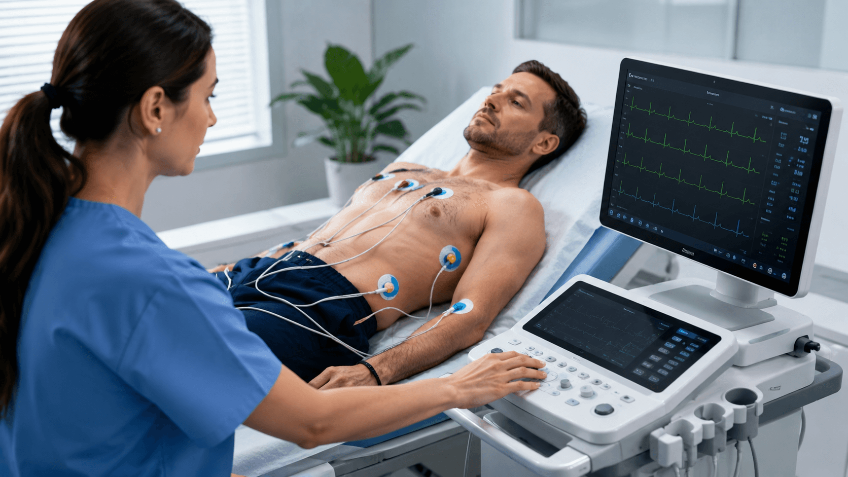
A patient presents with symptoms compatible with acute coronary syndrome. The first electrocardiogram does not show persistent ST-segment elevation that meets the conventional threshold for a working diagnosis of ST-elevation myocardial infarction.
That observation is important. It is not equivalent to showing that every coronary artery is patent.
The distinction matters in the other direction as well. An ECG can satisfy a pattern used to activate a ST-elevation pathway while subsequent investigation identifies no acute culprit occlusion. The ECG finding is real; the anatomical interpretation attached to it may still be wrong.
Three quantities must therefore remain separate:
- The observed electrical pattern: what the ECG records at the body surface.
- The underlying coronary and myocardial state: whether there is acute culprit-vessel occlusion, severe flow limitation, ischaemia and evolving injury.
- The clinical action: whether the total evidence supports immediate angiography, another time-sensitive pathway or continued diagnostic evaluation.
These quantities are related, but none is a synonym for either of the others. Treating them as interchangeable creates two symmetric errors: failure to escalate an acute occlusion that does not produce the expected pattern, and unnecessary emergency activation when another condition produces a similar pattern.
The engineering problem is not to dismiss ST elevation. It is to understand exactly what that measurement can establish, what it cannot establish alone and how a new decision-support system would have to be validated before it could change care.
ST elevation is a measurement-defined phenotype
An ECG records voltage differences between electrodes placed on the body. Each lead is a particular view of the aggregate electrical activity projected to the body surface. ST-segment displacement is therefore an observed electrical phenotype, not a direct image of a coronary lumen.
In the appropriate clinical context, persistent ST elevation is a highly consequential sign of ongoing acute coronary occlusion. It supports a time-critical reperfusion pathway because delay can increase irreversible myocardial injury. Its value comes from this clinically established association and from the speed and availability of the ECG.
Association does not create identity. The visible pattern depends on more than whether thrombus is present in a coronary artery. It is affected by the location and extent of ischaemia, the direction of the resulting electrical changes relative to the recorded leads, the timing of the ECG, collateral flow, prior infarction, ventricular hypertrophy, bundle-branch block, ventricular pacing, pericardial or myocardial inflammation, electrolyte disturbances and technical quality.
That list should not be read as an invitation to explain away an abnormal ECG. It defines why the ECG must be interpreted as evidence about a latent physiological process rather than as direct anatomical ground truth.
The time axis is equally important. Coronary flow and myocardial electrical behaviour can evolve between symptom onset, the first ECG, serial ECGs, treatment and angiography. A single tracing is a sample taken from that trajectory. A later angiogram is a different measurement taken at a different point in the same evolving event.
This is why the relevant question is not simply:
Does this ECG cross a fixed ST threshold?
It is:
Given the symptoms, timing, ECG morphology and evolution, haemodynamic state, biomarkers and other available evidence, how strongly does this presentation support ongoing acute coronary occlusion or another condition requiring immediate invasive management?
The second question is harder, but it corresponds to the clinical decision that actually has to be made.
Occlusion, infarction and the treatment pathway are different labels
Terminology can hide distinctions that the evidence requires.
Acute coronary occlusion is an anatomical and physiological claim about a culprit artery and coronary blood flow. Angiography can provide the strongest routinely available evidence about that state, but the definition still requires choices: complete versus near-complete obstruction, Thrombolysis in Myocardial Infarction flow grade, evidence that the lesion is acute rather than chronic, and whether flow after spontaneous or treatment-associated reperfusion counts.
Myocardial infarction is a clinical diagnosis of acute myocardial injury in the setting of myocardial ischaemia. The Fourth Universal Definition requires a rise and/or fall in cardiac troponin with at least one value above the 99th-percentile upper reference limit, together with specified evidence of acute ischaemia. Infarction is therefore not defined by coronary occlusion alone, and an elevated troponin alone is not necessarily infarction. Thygesen et al., Journal of the American College of Cardiology (2018)
STEMI and NSTE-ACS are working diagnoses used to organize urgent evaluation and treatment. They are not literal angiographic descriptions. The 2023 European Society of Cardiology guideline explicitly notes that myocardial infarction will not be the final diagnosis in every patient with a working diagnosis of STEMI. It also states that some cases warrant prompt triage for immediate reperfusion despite the absence of ST elevation. ESC Guidelines for the management of acute coronary syndromes (2023)
Occlusion myocardial infarction, or OMI, is a research and clinical framing intended to focus attention on infarction caused by an acute culprit lesion for which immediate reperfusion may be important, including presentations that do not meet conventional STEMI criteria. It is not one universally standardized outcome label, and it has not replaced STEMI/NSTE-ACS terminology in major acute-coronary-syndrome guidelines.
Those qualifications are not semantic caution. They determine which patients enter a study, which cases count as positive and whether two reported performance results are meaningfully comparable.
Current guidelines do not rely on one threshold alone
A serious account of this problem should not caricature standard care as “ST elevation or nothing.”
The 2023 ESC guideline uses the initial 12-lead ECG to place suspected acute coronary syndrome into two working pathways: persistent ST elevation or an equivalent pattern, and suspected non-ST-elevation acute coronary syndrome. It recommends that the ECG be obtained and interpreted promptly, repeated when necessary and considered alongside clinical context and patient stability.
It also identifies circumstances in which an ECG that does not meet the standard persistent-ST-elevation pattern may still support immediate management. These include recognized equivalent patterns, ongoing symptoms and findings suggestive of posterior or right-ventricular ischaemia. In a patient with high clinical suspicion of ongoing ischaemia, bundle-branch block or a paced rhythm can prevent reliable assessment of ST elevation. The guideline further recommends an immediate invasive strategy for very-high-risk NSTE-ACS, including presentations with haemodynamic instability, recurrent or refractory chest pain, life-threatening arrhythmia, acute heart failure caused by ongoing ischaemia, mechanical complications or recurrent dynamic ECG changes. ESC Guidelines for the management of acute coronary syndromes (2023)
This has two implications.
First, an ECG decision-support system must be evaluated against the actual guideline-informed workflow and qualified clinicians, not against a deliberately weakened baseline containing only one numerical cut-off.
Second, improved recognition of an anatomical target on retrospective data does not by itself establish that a new system improves the clinical treatment decision. The current pathway already combines electrical, symptomatic, haemodynamic and biomarker evidence. A new method has to demonstrate incremental value at the point where a real decision is made.
“OMI” is not one ground-truth definition
Three major ECG studies illustrate the definitional problem.
In a 2023 multi-site prospective observational study, Al-Zaiti and colleagues defined OMI primarily by an angiographic culprit lesion with TIMI flow grade 0 or 1. The definition also included a restricted category with TIMI 2 flow, severe stenosis and a high peak troponin. The investigators developed their model in 4,026 patients presenting with chest pain and evaluated it in two external cohorts, for a total of 7,313 patients. Patients with a confirmed prehospital STEMI were excluded, so the study addresses recognition among presentations not already captured by that overt prehospital pattern. Al-Zaiti et al., Nature Medicine (2023)
In a 2024 international retrospective evaluation, Herman and colleagues used a different composite. OMI required symptoms and troponin findings consistent with myocardial infarction plus either angiographic TIMI 0–1 flow or TIMI 2–3 flow with emergent or urgent revascularization. The latter branch partly incorporates a treatment decision into the target label. Herman et al., European Heart Journal – Digital Health (2024)
In a 2026 Swedish emergency-care study, Gustafsson and colleagues used a stricter anatomical primary outcome: newly formed complete or near-total culprit occlusion recorded at catheterization, operationalized as TIMI 0 flow for the principal OMI definition, with acute rather than chronic status determined from the angiographic procedure. Gustafsson et al., Nature Communications (2026)
These labels overlap. They are not identical.
A model evaluated against strict TIMI 0 occlusion is answering a narrower question than one evaluated against a composite that can include residual flow and urgent revascularization. A label containing troponin magnitude depends on the assay, sampling schedule and time since symptom onset. A label containing the decision to revascularize can inherit local practice and clinician judgement. A label assigned at catheterization can differ from the vessel state at the time of the prehospital ECG.
Accordingly, “OMI sensitivity” has no complete meaning unless the report also states:
- the target definition;
- when the ECG was acquired relative to symptoms and treatment;
- when and how angiography was performed;
- how acute and chronic occlusion were distinguished;
- how cases without angiography were handled;
- which troponin assay and thresholds were used;
- whether revascularization decisions were part of the label; and
- which patients were excluded before evaluation.
Without those details, a high score may be precise about an outcome that is not the one a hospital intends to use.
What the published model studies actually show
The recent literature provides credible evidence that the 12-lead ECG contains information relevant to acute occlusion beyond a single threshold rule. It does not yet provide evidence that an autonomous model improves patient outcomes.
A prospective, multi-site chest-pain cohort
The 2023 Nature Medicine study reported 7,313 consecutive patients across its development and external-validation cohorts. OMI prevalence was 5.2% in the 4,026-patient derivation cohort and 6.4% across the 3,287-patient external cohorts.
In held-out development data, the model's area under the receiver-operating-characteristic curve was 0.91, compared with 0.79 for practising clinicians and 0.78 for a commercial ECG algorithm. In external validation, the reported area was 0.87. At the study's selected rule-in and rule-out thresholds, sensitivity was 0.86, specificity 0.98, positive predictive value 0.54 and negative predictive value 0.99.
These results are material because the population was not a balanced laboratory dataset and the model was evaluated at independent sites. The limitations remain material too. The study excluded presentations already identified as prehospital STEMI, cardiac arrest and ventricular tachyarrhythmia. The output did not direct clinical care. The reported estimate of improved reclassification is therefore a retrospective or observational decision-analysis result, not evidence of reduced mortality, smaller infarcts or faster safe reperfusion.
The false negatives are also instructive. The authors reported 28 false-negative cases at their selected operating point; low-voltage signals, noise or baseline wander, a benign-appearing tracing and previous myocardial infarction were common among them. Those errors identify operating conditions that require explicit validation. They do not support a blanket statement that the remaining missed cases are inherently undetectable.
An international retrospective comparison
The 2024 European Heart Journal – Digital Health study developed its model using 18,616 ECGs from 10,543 patients with suspected acute coronary syndrome. Its reported test set contained 3,254 ECGs from 2,222 patients, with an OMI prevalence of 21.6%.
The model achieved an area under the curve of 0.938, with sensitivity of 80.6% and specificity of 93.7% at the reported threshold. In that evaluation, conventional STEMI criteria had sensitivity of 32.5% and specificity of 97.7%, while expert interpretation had sensitivity of 73.0% and specificity of 95.7%.
The result supports the hypothesis that an ECG model can improve sensitivity for the study-defined target while retaining high specificity. It does not make the operating point universal. The OMI prevalence, composite reference definition and inclusion process affect predictive values and case difficulty. The study was retrospective, and several authors disclosed founder, employment or ownership relationships with the company developing the model. A declared conflict does not invalidate a result, but it increases the importance of independent replication and prospective evaluation.
Scale does not remove class imbalance or transfer risk
The 2026 Nature Communications study analysed 540,372 emergency ECGs associated with 465,471 visits by 225,824 patients. Its outcome data included 1,583 OMI events, 4,279 myocardial infarctions without OMI and a much larger control group. OMI therefore represented approximately 0.3% of the study ECGs.
The model achieved C-statistics of at least 0.95 for OMI in its internal random and temporal test sets. At a 5% false-positive rate, sensitivity was 0.87 in the Swedish emergency-department test sets. The authors also reported lower average precision in external testing, weaker performance in some clinically important conditions and no external dataset with complete culprit-vessel localization labels.
The dataset's scale is a strength. The low event prevalence is also a warning against reading discrimination as operational usefulness. Even a small false-positive fraction, applied to a very large non-OMI population, can produce many false escalations. The study itself concludes that randomized trials with patient-relevant outcomes are needed before clinical deployment.
Across all three studies, the defensible conclusion is bounded: ECG-based models can discriminate a declared OMI outcome in retrospective or observational cohorts, including independent cohorts. None of these studies proves that model-directed triage improves clinical outcomes or that a reported threshold transfers unchanged to a new care system.
AUROC is not the clinical objective
Area under the receiver-operating-characteristic curve measures how often a randomly selected positive case is ranked above a randomly selected negative case across all possible thresholds. It does not specify the threshold at which a system will operate, the number of false catheterization-laboratory activations it will produce or how many occlusions it will miss.
For a time-critical escalation system, at least six quantities have to be reported at a prespecified operating point:
- Sensitivity: the proportion of patients meeting the declared occlusion definition who trigger the output.
- Specificity: the proportion not meeting that definition who do not trigger it.
- Positive predictive value: the proportion of triggered outputs that meet the definition in the evaluated population.
- Negative predictive value: the proportion of non-triggered outputs that do not meet the definition in that population.
- Coverage: the proportion of eligible cases for which the system issues a determinate result rather than abstaining.
- Decision latency: the time from availability of the required data to an actionable output.
Predictive values change with prevalence. A threshold evaluated in a cohort where one in five patients has OMI will not retain the same positive predictive value in an operational stream where the condition is one in several hundred ECGs, even if sensitivity and specificity remain unchanged.
Average precision or a precision–recall curve is particularly informative for rare outcomes because it makes the trade-off between recovered positive cases and false alerts more visible than AUROC alone. Calibration is separately required: among patients assigned a probability near 0.20, approximately one in five should meet the prespecified outcome in a suitable validation sample. Good ranking does not guarantee good calibration.
The clinical comparison must also be defined at equal resource constraints. Comparing a model's sensitivity at one specificity with a clinician's sensitivity at another does not establish which approach produces the better workflow. A credible study should compare strategies at prespecified false-activation rates or use a decision analysis that explicitly values delayed reperfusion, unnecessary emergency angiography, alternative diagnoses and abstention.
Detecting occlusion and localizing a culprit are separate claims
“An acute occlusion is present” and “the culprit is in this coronary territory” are different outputs with different error structures.
The first may support escalation. The second may influence procedural preparation or the interpretation of additional evidence. A model can perform well on detection while confusing adjacent territories, especially where the electrical projection is less distinctive or anatomy varies.
The 2026 Swedish study illustrates the distinction. Its authors reported strong internal discrimination for OMI while identifying difficulty separating left-circumflex from right-coronary culprits. They also lacked external validation sets with the angiographic labels required to test localization fully.
It would therefore be incorrect to convert a validated detection claim into an unvalidated localization claim. Each output needs its own reference definition, error matrix, subgroup analysis and external test.
The failure cases define the product
A clinically responsible system cannot be specified only by its average performance. It must state what happens when the evidence is weak, conflicting or outside validation.
Relevant conditions include:
- left or right bundle-branch block;
- ventricular pacing;
- left-ventricular hypertrophy;
- prior myocardial infarction or chronic ST–T abnormalities;
- pericarditis and myocarditis;
- electrolyte disturbance;
- low signal amplitude, baseline wander and electrode artefact;
- ECG acquisition early or late in the event;
- spontaneous or treatment-associated reperfusion before angiography;
- acute versus chronic total occlusion;
- culprit territories represented by small case counts; and
- patients who never undergo angiography.
A single forced label conceals these distinctions. A professional decision-support output should identify whether the evidence is sufficient for the intended use, state its uncertainty, and preserve escalation when the clinical context is more concerning than the ECG result.
This does not mean displaying an unstructured list of model features. It means communicating the status of the claim:
- which clinical question the output addresses;
- whether it concerns occlusion detection, localization or both;
- which data were available and technically usable;
- whether the presentation falls within the validated population;
- what operating threshold was applied;
- what action the system supports, if any; and
- when repeat ECG, additional conventional leads, imaging or specialist assessment remains necessary under the clinical pathway.
The final action remains a clinical decision. A probability of occlusion is not itself an order for angiography, just as failure to cross a probability threshold is not proof that an artery is open.
A defensible validation programme
Any system intended to affect acute-coronary-syndrome triage should progress through evidence stages that answer different questions.
1. Define the intended use and target before analysis
The population, setting, user, input timing and action must be fixed. “Detect OMI” is insufficient. A valid protocol might instead specify decision support for adults with suspected ACS who have an interpretable pre-treatment 12-lead ECG and have not already met the institution's immediate invasive criteria.
The OMI endpoint must be predeclared, including angiographic flow, acute-versus-chronic adjudication, troponin requirements, handling of reperfused vessels and cases without angiography.
2. Use consecutive clinical cohorts
Case-control enrichment can estimate whether a signal exists, but it cannot directly establish predictive value or alert burden in practice. Evaluation should retain the natural prevalence, relevant alternative diagnoses and technically poor ECGs encountered in the intended workflow.
3. Prevent patient, site, device and time leakage
ECGs from the same patient must not cross development and test boundaries. Site-disjoint testing evaluates institutional transfer. Device-disjoint testing evaluates dependence on acquisition and preprocessing. Temporal testing evaluates changes in population and practice. An external threshold should remain prespecified; tuning it on the test site converts external validation into local model development.
4. Adjudicate the reference independently
The reference committee should be blinded to the investigational output and apply the declared definition consistently. Reports should give inter-reviewer agreement, unresolved cases and reasons for exclusion. Angiography, serial biomarkers, symptoms, treatment timing and clinical course should remain separately available so that disagreement about a composite endpoint can be audited.
5. Test the known hard subgroups
Performance should be reported for conduction abnormalities, pacing, ventricular hypertrophy, prior infarction, inflammatory mimics, low-quality recordings, symptom-to-ECG time and culprit vessel. Small subgroup denominators and confidence intervals must be visible. “No statistically significant difference” is not evidence of equivalence when the study is underpowered.
6. Run prospectively in silent mode
Before affecting care, the system should operate on live data without changing decisions. This stage tests data availability, latency, ECG versioning, repeated recordings, missingness, user eligibility and the frequency and causes of abstention.
7. Test clinical impact
Only a prospective controlled study can establish whether making the output visible changes care beneficially. Appropriate endpoints include time to angiography or reperfusion for adjudicated occlusion, missed or delayed cases, false emergency activations, complications, infarct size or cardiac function where justified, length of stay and patient-relevant outcomes. The study should also detect harmful over-reliance when the model disagrees with clinical evidence.
Randomized evaluation may be necessary for a claim of patient benefit. Retrospective reclassification cannot substitute for it.
What the public evidence proves—and what it does not
The public evidence supports four conclusions.
First, ST elevation is a valuable but indirect electrical sign, not an anatomical measurement. Second, acute occlusion can occur without the conventional persistent-ST-elevation phenotype, and an apparent STEMI pattern does not guarantee an acute culprit occlusion. Third, current guidelines already account for equivalent patterns, clinical instability and very-high-risk non-ST-elevation presentations. Fourth, multiple ECG-model studies have shown promising discrimination for explicitly defined OMI outcomes across large and, in some cases, external cohorts.
The evidence does not establish one universal OMI definition. It does not show that every angiographic occlusion is identifiable from one ECG. It does not prove that model probabilities are calibrated at a new hospital, that localization transfers across populations or that model-directed care improves outcomes. It does not establish the performance, safety, regulatory status or clinical readiness of any Mondren system.
The most accurate summary is therefore not that STEMI is obsolete. It is that the measurement, the target condition and the action should not be collapsed into a single label.
The Mondren perspective
At Mondren, acute coronary occlusion is a frontier research problem. Our interest is in systems that can make the evidential structure of a time-critical decision more explicit while remaining accountable for what the measurements cannot resolve.
Our public position is:
ST elevation should be treated as important evidence of acute coronary occlusion in the appropriate context, not as a literal observation of the artery. A credible system must distinguish the ECG phenotype, the underlying coronary state and the clinical action, and it must preserve uncertainty when those cannot be resolved safely.
This article reports no Mondren benchmark, clinical trial, product performance, regulatory clearance or patient-benefit claim. Every numerical result cited above comes from external public research.
Any future claim from us should identify the intended-use population, OMI definition, ECG timing, angiographic coverage, cohort prevalence, patient and site boundaries, operating threshold, sensitivity, specificity, predictive values, calibration, abstention rate, decision latency and hard-subgroup results. A clinical-impact claim should additionally specify how the output changed care and which patient-relevant outcomes were measured.
The problem definition and evidence standards can be public. Our internal representation, source reconstruction, lead-weighting logic, feature construction, sequence handling, uncertainty calculations and any measurement-selection logic remain proprietary.
The commercial implication
The commercial objective is not a higher retrospective score. It is safer and faster recognition of time-sensitive coronary occlusion without an unacceptable increase in emergency activations or false reassurance.
For an emergency medical service, that claim depends on prehospital acquisition quality, transmission, latency and the receiving system's pathway. For an emergency department, it depends on how the output interacts with serial ECGs, biomarkers, haemodynamics, specialist review and access to catheterization. For an ECG or clinical-software manufacturer, it also depends on device variation, human factors, auditability, fail-safe behaviour and the regulatory definition of intended use.
The appropriate initial boundary may be an escalation and review aid rather than autonomous diagnosis or cath-lab activation. That boundary is not a marketing qualification. It determines the harm model, the validation endpoints and the evidence burden.
A useful system would identify additional high-risk cases early enough to change the diagnostic pathway. A trustworthy system would also identify when its ECG evidence is insufficient, when the case is outside validation and when other clinical findings must override its result.
ST elevation and coronary occlusion are connected by physiology. They are not the same object. Preserving that distinction is the starting point for better measurement, better validation and safer decision support.
This article discusses cardiovascular research and engineering evaluation. It is not medical advice and does not make a claim of clinical safety, efficacy, regulatory clearance or patient benefit.